 To main content
To navigation
To main content
To navigation
Breakthrough in metabolic disease research using infrared free-electron laser
Researchers from HFML-FELIX have successfully identified molecules related to two rare genetic disorders, directly from brain tissue using a powerful infrared free-electron laser. This new analytical technique contributes to a better understanding of these diseases and may lead to faster diagnoses and more effective treatments in the future.
All cells in our body are constantly making and breaking down substances. This is necessary, among other things, for processing food, maintaining sufficient energy, and supporting growth. We call this process metabolism. In the case of an inherent metabolic disorder, something disturbs this process. One of the enzymes needed to convert one substance into another cannot be made properly or cannot be made at all, due to an error in a gene. This can interfere with our body’s processing of certain substances, resulting in an accumulation of harmful molecules or lack of essential ones, often with severe effects on the brain.
In his experiments, HFML-FELIX researcher Jelle Schuurman has looked at two metabolic diseases – pyridoxine-dependent epilepsy and glutaric aciduria type 1 – that prevent a specific substance from being processed properly, namely the substance lysine. The consequences of this disturbed process are particularly visible in the brain. Therefore, researchers want to understand this process as complete as possible. Which substances accumulate, for example? And by extension: what do we see when we apply certain treatments? What changes then?
Not only mass, but also structure
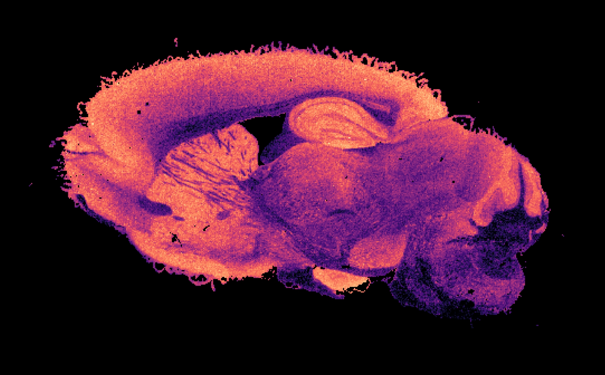
Hospital and research laboratories can already tell quite a lot about which molecules are present in a complex fluid like a blood sample. They do this by using mass spectrometry, among other methods. This involves measuring the mass of molecules, looking at how many of those different masses are present. In the case of a piece of tissue, mass spectrometry imaging is used to tell exactly where these molecules are located. You can see the result of this in the image above this article. However, this still doesn’t tell you everything. Two molecules can weigh the same but have completely different structures and functions. Therefore, it is also necessary to say something about the structure of each molecule. Only then do you know for certain which one you are dealing with.
To find out more about this, Schuurman used the powerful infrared free-electron laser FELIX. With this laser, it is possible to make the molecule you are studying react in a certain way. Molecules each have unique frequencies at which they vibrate. This even varies for different parts of the same molecule. If you send light with different frequencies through a molecule, it reacts to a matching frequency by vibrating. With a powerful laser, you can even make them vibrate so much that they break. By looking at which frequencies cause the molecule to fall apart, the structure can subsequently be determined.
Completely New Technique
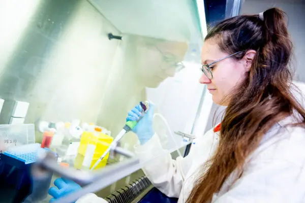
This is a valuable existing technique, but Schuurman and his colleagues looked at whether they could push this even further. The existing approach works very well for bodily fluids. But what if you want to know which molecules are in a solid substance? For example, when you want to examine the brain, as with the aforementioned metabolic diseases. For this, naturally, no samples from living patients are used, but samples from mice with the same genetic abnormality. In some cases, artificially grown organs with genetic material from the patient are available, or tissue from deceased patients.
In this research, molecules in a piece of mouse tissue were first mapped using a UV laser. Then FELIX was used to identify molecules most closely correlated to the studied genetic disorders. Nowhere else in the world has that been achieved in this way before, Schuurman explains. ‘Being able to identify molecules directly from biological tissue, in low concentrations and without having to do an extraction or purification, is completely new. We did not get it right immediately either, but we are very enthusiastic that we eventually succeeded. We are getting a lot of positive reactions from colleagues in the research field.’
Faster diagnoses, more effective treatments
That we made this analytical technique work is the most important discovery within this research, but it also gives researchers more knowledge about these two specific metabolic diseases. Ultimately, this technique can also be used to study what changes in the brain when different treatments are used. Which molecules do we find then? Because it would be possible to genetically disable the disrupted pathways – like the processing of lysine – as a treatment, you want to first be able to study what the consequences of such a treatment would be.
The direct impact of this breakthrough therefore lies particularly in biomedical research. It not only accelerates fundamental research into disease mechanisms but also provides a powerful instrument for developing new diagnostic methods and for evaluating treatments. Schuurman and his team are therefore very pleased with this achievement. ‘We are incredibly enthusiastic about the possibilities this offers. This is an essential step that enables us to better understand diseases, diagnose them faster, and ultimately treat them more effectively.’
You can find the paper here:
Structure Elucidation for MALDI Mass Spectrometry Imaging Using Infrared Ion Spectroscopy
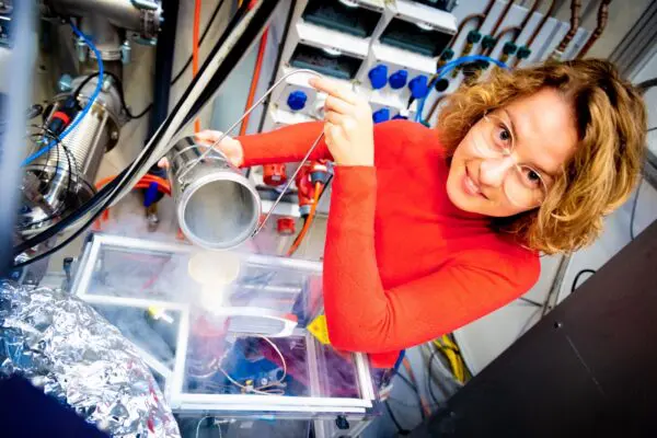
Research contact
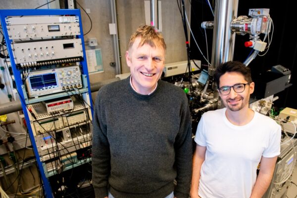
Jelle Schuurman
More info about Jelle

Dr. Jonathan Martens
More info about Jonathan

Other news
Why researchers want to levitate living cells in high magnetic fields
‘When I finish an experiment, it is often not the end of something, but the beginning.’

HFML-FELIX at NWO Physics 2026: Exploring Matter Under Extreme Conditions

When electrons sing in harmony

Happy holidays from all of us!
HFML-FELIX researcher Olga Duda wins NWO Poster Award second year in a row
How a sunscreen molecule that encounters metal ions can absorb light in a very different way

Promising new technologies for researching inherited metabolic diseases

First four projects to receive funding out of the new Incentive Fund

Chemists find clues to the origins of iconic ‘football molecule’ in space

Apply for research time before 15 November

Radboudumc joins HFML-FELIX as an official partner to advance metabolic disease research
Exciting research position in newly granted European project!

Unraveling the secrets of space ice
Fingerprints of important interstellar molecules determined

Registration open for the ‘Astrochemistry: From Quantum to Cosmos’-meeting
A new, creative approach to switching magnetization
HFML-FELIX researchers win prize for molecules that stimulate crop growth
A successful 2025 HFML-FELIX User Meeting
Shedding light on a promising organic catalyst