 To main content
To navigation
To main content
To navigation
Why researchers want to levitate living cells in high magnetic fields
For years researchers have been working on growing organs from living cells in the lab. Having this lab grown tissue, could one day be a solution for the shortage of donor organs. This line of research already had some great successes along the way. However, the exact process that makes cells grow together to form tissue and organs, remains very difficult to recreate in a lab setting.
Of course tissue in our body does not grow flat, like it would in a petri dish in the lab – to create an organ, cells need to grow in 3D. In the lab you can use a scaffold to help them grow into the right shape, but with the scaffold degrading, the shape and function of the organ might change over time. A novel idea is to apply external physical fields, such as a magnetic field, to levitate cells and steer them into 3D structures.
Levitating cells in a magnetic field
Not so long ago researchers came to HFML-FELIX to see whether magnetic levitation could be used to guide cells to grow in 3D-shapes and then hold them in that shape until the construction was strong enough to ‘stand’ on its own. To do so, the cells need to be in a very specific magnetic salt solution. This salt in high concentrations however, is very toxic for the cells. And if you use low quantities, you need very powerful magnets to get the same floating effect.
That is why the researchers ended up at HFML-FELIX, in Nijmegen, where such magnetic fields can be made in a research setting. What you eventually want to study, if you successfully get the cells to levitate, is: can they assembly into different shapes? And what influences the shape you get? Can you play with this by, for instance, tuning the magnetic field, or moving the sample in the set up?
Building a new microscope
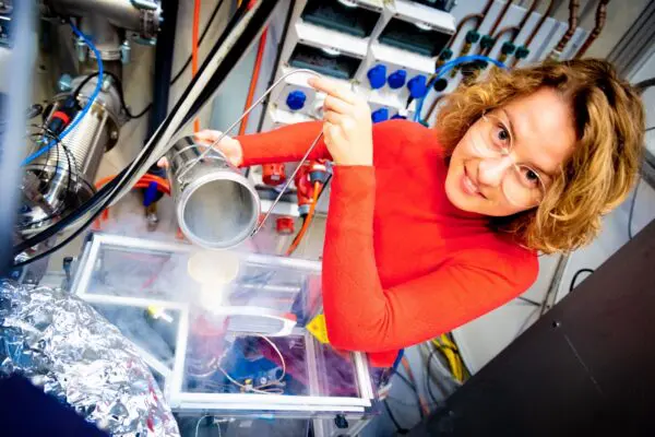
Today HFML-FELIX researcher Hannah Willenberg is working on answering these questions with new experiments. Not only is she looking at levitating cells and creating new assembly shapes, but at the same time she is also studying what the magnetic field does to the cells. To look at this they had to build their own confocal microscope that is compatible with the magnet and can create images from a meter distance away from the actual sample. Because that is how far down the samples have to be to get them in the right spot in the magnet for the highest magnetic field.
‘A huge part of my PhD-project was building this research setup from scratch’, Willenberg says. ‘The microscope has to be mounted close to the magnet, without touching the magnet to omit vibrations caused by the magnet cooling. As you can imagine, vibrations will make an image blurry, and we want pictures that are as sharp as possible. The good thing is: we now have this microscope working, and that means we can use it for other research questions too. It is suitable for any experiment in which you want to look at a small sample at room temperature in the centre of a magnetic field or under microgravity conditions.’
How to see them
To actually see what the magnetic field does to a living cell, you need more than just the microscope. You have to find a way to see the changes a cell goes through. This can be done by giving certain parts of the cell a fluorescent label, which you can see when you make an image with the microscope. When that part of the cell breaks down, or changes its behaviour, you can see these changes in the images. These are important experiments to do if you actually want to use magnetic fields to create organ tissue. You need to make sure the magnetic field does not harm the cells.
Levitating living cells
One of the other next steps is to levitate cells in the magnet to see whether we can form new shapes and layered structures. ‘Even though we use magnetic fields in health settings already – think of MRI-scanners in hospitals – there is a lot we don’t know about the effect of high magnetic fields on living cells’, Willenberg explains. ‘There is a lot of research we still need to do. And I hope my experiments can contribute to our knowledge about this subject.’
Credit picture: Gideon Laureijs
Research contact
Hannah Willenberg
More info about Hannah

Other news
A better understanding of the structure of steroid hormones

Two HFML-FELIX projects receive funding for modernizing research infrastructure

Sandra Brünken receives Special Review Lecturer Award

Finally, we know more about the elusive magnetic properties of small groups of atoms
Four new projects receive funding out of the HFML-FELIX Incentive Fund

Apply for research time before 15 May

Holland High Tech funding for detection and identification of chemical unknowns

‘When I finish an experiment, it is often not the end of something, but the beginning.’

HFML-FELIX at NWO Physics 2026: Exploring Matter Under Extreme Conditions

When electrons sing in harmony

Happy holidays from all of us!
HFML-FELIX researcher Olga Duda wins NWO Poster Award second year in a row

How a sunscreen molecule that encounters metal ions can absorb light in a very different way

Promising new technologies for researching inherited metabolic diseases

First four projects to receive funding out of the new Incentive Fund
Chemists find clues to the origins of iconic ‘football molecule’ in space

Apply for research time before 15 November

Radboudumc joins HFML-FELIX as an official partner to advance metabolic disease research
Exciting research position in newly granted European project!

Unraveling the secrets of space ice